
Have you ever wondered how it's possible to know which rib you are scanning without being able to see all the ribs in one image?
Start your examination by placing your transducer in a Sagittal plane on the body, short axis to the medial aspect of the palpable clavicle.
💡The clavicle is an easy landmark to begin because you visibly see the way the bone runs and place your transducer perpendicular to it. If you start your exam too far lateral you will miss the first rib and your count will be off by 1.
💡 When you visualize the first rib just inferior to the clavicle you can begin counting from 1-12.💡
1️⃣Move the transducer from
superior to inferior while
gradually gliding laterally as
you scan inferior
2️⃣When you are scanning along the medial aspect of the rib cage, you are at the level of the costal cartilage
3️⃣You must move laterally as you scan to see all 12 ribs. The true ribs are 1-7, the false ribs are 8-10, and the floating ribs are 11-12
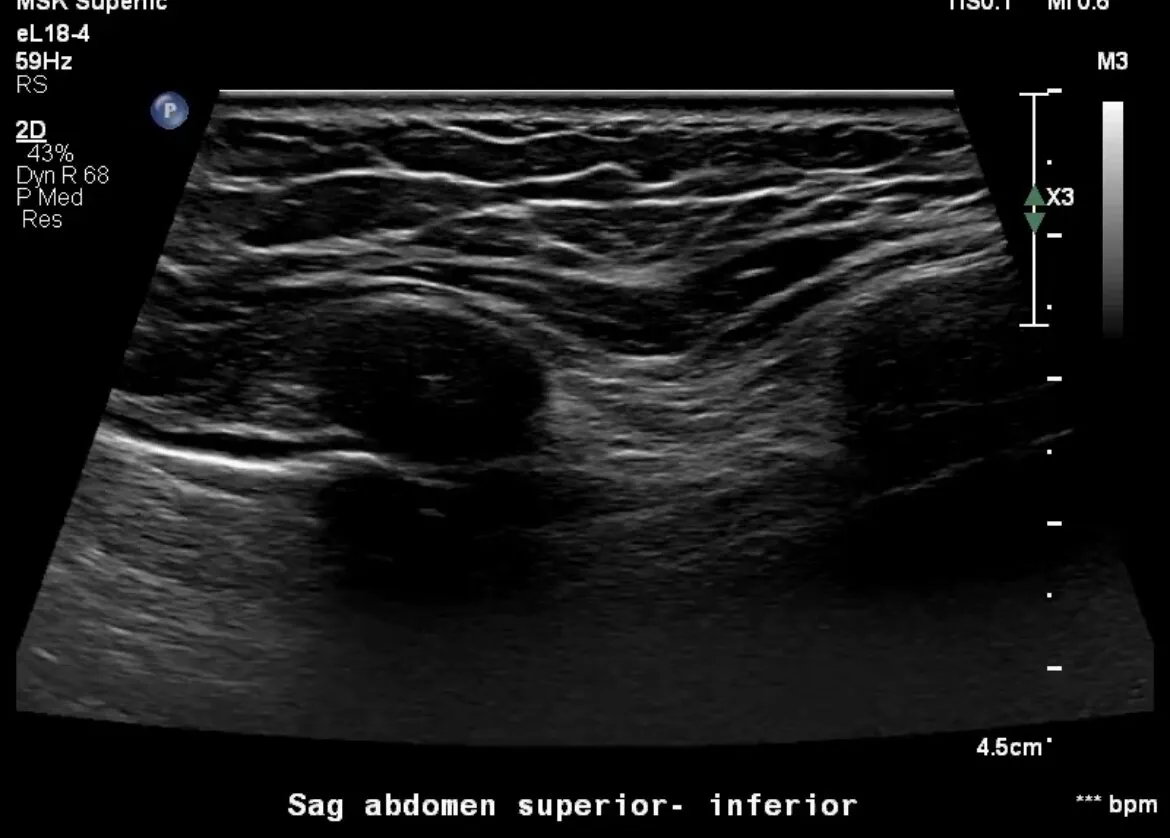
💡The rib appears hypoechoic at the level of the costal cartilage and transitions into a hyperechoic ossified bone laterally.💡
To begin evaluation of a single rib in the long axis, place the transducer in a transverse plane on the body over the rib of interest and do a sweep from medial to lateral. Start at the level of the bony sternum and sweep laterally until the costal cartilage transitions into a bony rib.
💡 You must keep elongating the rib while moving upward and lateral to fully visualize it.
❗️It is easier to count the ribs in the short axis when you are in a sagittal plane of the body to get an accurate count, but you must visualize the rib in both planes to know which segment of the rib you are looking at❗️
👏Now you are ready to label the ribs properly and examine them for pathology!
To watch the video Click Here