Log In
Log In
BLOG
What are 6 sonographic features of tennis elbow?

What are 6 sonographic features of tennis elbow?

⭐️ In light of the amazing career of Serena Williams, this week I am serving up 6 sonographic features that represent the classic ultrasound findings associated with tennis elbow:


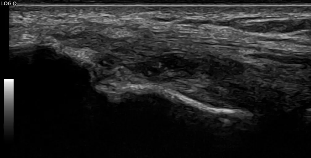
🎾Osteophytes on the lateral epicondyle


🎾Diffuse or focal hypoechoic, heterogeneous, and/or thickening of the common extensor tendon indicative of tendinopathy


🎾Focal hypoechoic or anechoic disruption of tendon fibers due to varying degrees of tearing


🎾Calcifications present within the common extensor tendon


🎾Hypermia present with the use of power doppler


🎾Secondary thickening of the underlying Radial Collateral Ligament


Click here to watch video

0 Comments
Add Comment

WARNING: DEMO IN SESSION! DO NOT CLOSE BROWSER.

 ULTRASCAN REMOTE

FULL DEMO

APPLY FOR REMOTE TRAINING

Dynamic Ultrasound of UCL

There is slight widening of the joint space with valgus stress.


BECOME A FRIEND

Copyright 2019+ learnmsksono.com | All rights reserved

© This site is not a part of the Facebook website or Facebook Inc. Additionally, This site is NOT endorsed by Facebook in any way. FACEBOOK is a trademark of FACEBOOK, Inc.

Powered by Liquid Funnels

Your cart is empty Continue
Shopping Cart
Subtotal:
Discount 
Discount 
View Details
- +
Sold Out